
By Stephen Bunting, Specialist MSK Physiotherapist
Can a disc really “slip” out of place? It’s one of the most common phrases I hear in clinic, usually said with a fair bit of worry attached to it — and it’s also one of the most misunderstood terms in the whole of musculoskeletal medicine.
In this article, I’ll explain what a spinal disc actually is, what can genuinely go wrong with one, and why the term “slipped disc” is outdated and inaccurate. I’ll also cover how to tell when a disc problem is actually significant, and when you do and don’t need a scan.
The information is also presented in video format below.
What Do Spinal Discs Actually Do?
You have 23 discs in total, sitting between the vertebrae of your spine. Their main job is to act as shock absorbers, cushioning the vertebrae from impact and spreading load through the spinal column. They also contribute to spinal movement, and help hold each vertebra securely to its neighbour, a bit like a ligament.
Each disc has two distinct parts: an inner part called the nucleus, which is a jelly-like substance, and an outer fibrous part called the annulus — a bit like a jam doughnut. As force is transmitted through the spine, the jelly-like nucleus gets squeezed and spreads out, but is contained by the tough fibrous annulus, allowing the disc to absorb and dissipate load.
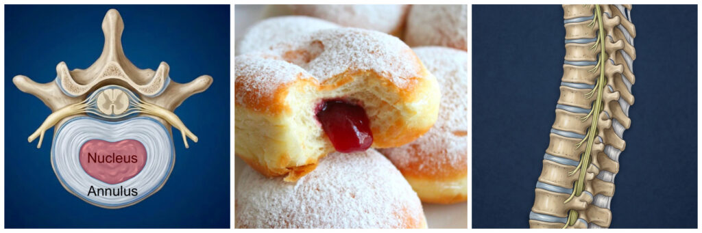
What the anatomy books don’t tell you is that your discs only look like this in your teens and early twenties. After that, discs start to change. The nucleus loses water and shrinks, and the disc loses height and gets thinner as the years progress. Collectively, as all 23 discs shrink with age, this is the main reason people lose height in later life — and it’s entirely normal, age-related change. So when you read “disc degeneration” or “disc desiccation” on a scan report, that’s simply describing this process: the disc has lost some water content and some height over time, much like grey hair or wrinkles. Most people in their 70s will have very little water left in most of their discs, which will be essentially all annulus with little to no nucleus remaining.
What Can Go Wrong With a Disc?
Now that we know what a normal disc looks like, let’s go through the various things that can happen to it, and the terms you’ll commonly see used on an MRI report.
Disc Bulges
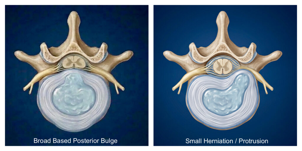
Discs are designed to bulge to some degree when the spine is active and under load, and sensible activity and exercise actually help maintain healthy discs. But due to the demands of modern life — particularly long periods of sitting, being overweight, or an inactive lifestyle — the annulus can weaken and stretch, and the disc can start to bulge outwards, a bit like a deflated car tyre. This is often reported on a scan as a “broad-based disc bulge.” Because the annulus is weakest at the back, the disc commonly bulges backwards, which you might see reported as a “posterior disc bulge” — this is the most common disc-related finding of all.
Disc Herniations and Protrusions
Sometimes a bulge is very localised, involving only a small portion of the disc surface. This is called a disc herniation. Small herniations are very common, caused when a section of the outer annulus weakens and the inner nucleus pushes against it, creating a very localised bulge — a bit like a blister. These are often referred to on a scan as a “disc protrusion.” Bulges and small protrusions are extremely common and usually don’t cause any problems, and they will often reduce over time naturally, either as the annulus heals or as the water in the nucleus is gradually reabsorbed as part of normal ageing.
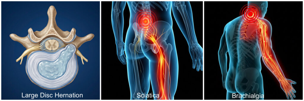
Larger herniations can cause symptoms though, because they can interfere with the nerve root that sits right next to the disc. Classic symptoms of nerve root irritation are pain, and sometimes tingling or numbness, travelling down the arm or leg rather than staying at the spine. In the lower back, this is what’s commonly called sciatica. In the neck, the equivalent term is brachialgia, or more correctly, cervical radiculopathy — the same kind of nerve root irritation, just affecting the arm instead of the leg.
Link to Sciatica (Lumbar Radiculopathy) article
Link to Cervical Radiculopathy article
Annular Tears and Disc Extrusions
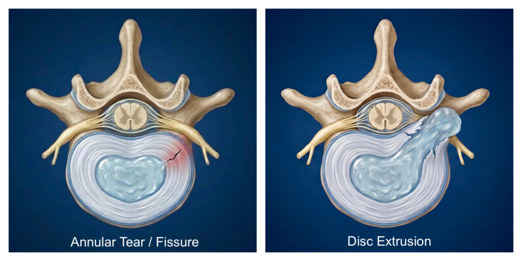
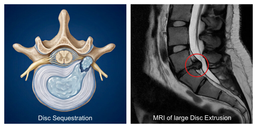
All of the disc bulges described so far are contained, meaning the outer surface of the annulus is still intact and the nucleus remains within it. But in some cases, the annulus develops small cracks, which show up on a scan as an “annular fissure” or “annular tear.” Small annular tears are really common, especially in older discs, and are usually just part of the normal ageing process. Occasionally, though, a tear can penetrate right through the deeper part of the annulus, reaching the nucleus, and create a track through which the nucleus can squeeze out — a bit like toothpaste squeezing out of a tube. The correct term for this is a disc extrusion, though some radiologists still use the older term “disc prolapse,” or may simply call it a large herniation — the key feature is that the nucleus is now outside the annulus. An extrusion is an uncontained, or ruptured, disc herniation, and it’s considerably more likely to cause nerve root symptoms than the contained bulges and protrusions described above.
Disc Sequestration
The final disc-related term you might see on a report is a disc sequestration. This is where a fragment of extruded nucleus has separated entirely from the main disc and is now floating freely in the spinal canal, again often causing nerve root symptoms.
It’s worth saying that if you do have nerve root pain in the arm or leg from one of these disc problems, the odds are generally good that it will improve over a few months. The body can heal and reabsorb herniated disc material even with extrusions and sequestrations, and nerves can adapt to cope with pressure from disc material — though not always. Larger herniations and more severely compressed nerve roots are less likely to settle on their own than smaller ones, and surgery is sometimes needed, particularly if symptoms are severe or haven’t improved after three or four months, so it’s worth seeing a spinal surgeon if you’re concerned.
So, Can a Disc Really “Slip”?
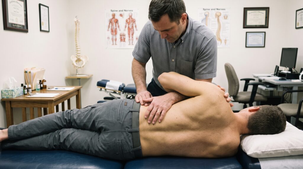
Medical professionals generally dislike the term “slipped disc,” because it’s old-fashioned and inaccurate. The people who tend to still use it are often the ones claiming they can manipulate your “slipped disc” back into place using spinal manipulation or a similar technique.
Hopefully you can now see how unlikely that claim really is. Imagine trying to put toothpaste back into the tube by moving the tube around in different directions — it simply isn’t going to happen. Discs don’t slip anywhere. They bulge, herniate, prolapse and extrude, but they don’t slip, and they can’t be manipulated back into place. It’s a term that persists largely because it’s useful for selling the idea that something can be “put back” for you, which isn’t an accurate description of what’s actually happening in the tissue.
Another thing I commonly hear about is people being told that their X-ray has shown a disc prolapse or a compressed nerve. You simply can’t see discs or nerves on an X-ray — only bone. An X-ray can show the space between vertebrae where a disc sits, and if that space looks narrow, you might assume the disc has thinned for some reason, but you can’t tell from an X-ray alone whether that’s just normal age-related degeneration or an actual herniation, or whether a nerve is being compressed or not.
X-rays can show the small holes — the intervertebral foramen — that nerves use to exit the spine, and if these look narrow, you might assume a nerve is being compressed, but again, you simply don’t know from an X-ray whether that’s actually affecting the nerve. To properly see a disc or a nerve, you need an MRI scan.
What About Older Discs?
Everything described above tends to happen in younger or middle-aged discs, where there’s still some nucleus left to bulge or herniate. Older discs behave differently. As the nucleus dries out, the disc becomes less efficient at absorbing load, so it tends to flatten, widen and sag slightly — much like other parts of the body as we age. This can still be reported as a “bulge” on an MRI, but it’s really a degenerative bulge, and it’s an extremely common finding in older age. It’s also what leads to the gradual disc thinning and height loss that happens in almost everyone by the time they reach their 70s.
Do You Need an MRI Scan?
Most people with back pain won’t need an MRI scan, because the result usually won’t change the course of treatment. This is because a large body of research shows that many people with no back or nerve pain at all have disc degeneration and arthritis visible on their scans. A finding of disc degeneration or a disc bulge on your report might be entirely irrelevant to your symptoms.
A scan becomes necessary when your symptoms suggest something more significant is going on — for example, persistent radiating pain, numbness or weakness in an arm or leg suggesting nerve root compression, or if you have any red flag symptoms such as severe or worsening night pain, sciatica alongside changes in bladder or bowel function or numbness around the genital area, fever, a history of cancer, a visible spinal deformity, or back pain that started after a serious accident or injury. If any of that applies to you, please do get checked out urgently.
For most people, though, the changes seen on a scan are simply the typical changes we see in almost everyone as they age. In nearly 30 years of clinical practice, I’ve never once seen a scan report in someone over 30 that essentially says “perfectly normal spine.” If you’ve already had an MRI and want to understand what the terminology on your own report actually means, I’ve covered that in detail in a separate article.
If you have had an MRI scan and want to know how to interpret the information, then the video below explains to read and understand your scan report.
Summary
Spinal discs are shock-absorbing structures that naturally change with age — thinning, losing water, and sometimes bulging or herniating. These changes are described using terms like degeneration, bulge, protrusion, herniation, extrusion and sequestration, and the vast majority are a normal part of ageing rather than a sign of serious damage.
What discs don’t do is “slip.” They can’t be manipulated back into place, and anyone claiming otherwise is describing something that simply isn’t possible with the anatomy involved. Most back pain settles without needing a scan at all, and a scan is really only necessary when your symptoms point to something more significant, or when red flag symptoms are present.
This article is for general information only and does not replace individual assessment. If you’re concerned about your symptoms, please consult a qualified healthcare professional. See our full medical disclaimer.