
By Stephen Bunting, Specialist MSK Physiotherapist
Kienböck’s disease is a relatively uncommon condition that affects one of the small bones in the wrist called the lunate. If you’ve recently been diagnosed, you’ve probably come across terms like bone death or avascular necrosis, which can sound alarming. While it is a potentially serious condition, understanding what it is, why it happens, and how it can be treated can make it much less daunting.
This article explains what Kienböck’s disease is, what causes it, the symptoms to look out for, how it is diagnosed, and the treatment options that are available.
The information is also available in video format below.
What is Kienböck’s Disease?
Kienböck’s disease is a condition in which the blood supply to the lunate bone becomes reduced or disrupted. The lunate is one of the eight small carpal bones that make up the wrist and sits near the centre of the joint, where it plays an important role in wrist movement and transferring forces between the hand and forearm.
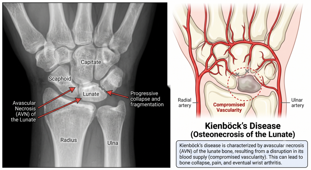
Like every other bone in the body, the lunate relies on a healthy blood supply to provide oxygen and nutrients. When this blood supply is interrupted, the bone tissue begins to weaken. Over time, the bone can become damaged, flatten, crack, or even collapse.
This process is known as avascular necrosis, which simply means damage to bone caused by a loss of blood supply.
As the condition progresses, the shape of the lunate may change, altering the mechanics of the wrist. In some cases, this can eventually lead to arthritis and ongoing wrist pain.
What Are the Symptoms?
The symptoms of Kienböck’s disease vary depending on how advanced the condition has become.
The most common symptom is pain in the middle of the wrist. This pain is often made worse by gripping, lifting, pushing through the hand, or activities that place weight on the wrist.
Other symptoms may include:
- Swelling around the wrist
- Stiffness
- Reduced grip strength
- Loss of wrist movement
- Difficulty performing manual tasks
Many people initially think they have simply sprained their wrist because the symptoms can develop gradually and feel very similar to a persistent wrist strain that never completely settles.
Why Does Kienböck’s Disease Happen?
One of the most common questions people ask is, “Why have I developed this?”
Unfortunately, there isn’t always one clear answer.
In many people, Kienböck’s disease is thought to develop because of a combination of factors rather than a single cause.
Reduced Blood Supply
The underlying problem is a loss of blood supply to the lunate bone.
Some people naturally have fewer blood vessels supplying the lunate than others. If that limited blood supply becomes disrupted, the bone may be more vulnerable to damage.
Wrist Anatomy
Another recognised risk factor is something known as negative ulnar variance.
This means the ulna—the smaller forearm bone on the little finger side—is slightly shorter than the radius.
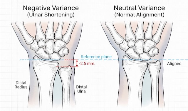
Researchers believe this may increase the forces passing through the lunate, placing it under greater mechanical stress over many years.
However, it is important to remember that many people have negative ulnar variance and never develop Kienböck’s disease, so this is unlikely to be the sole cause.
Previous Injury or Repetitive Loading
Some people notice symptoms following:
- A fall onto an outstretched hand
- Repetitive heavy manual work
- Regular exposure to vibrating tools
- Sports that repeatedly load the wrist
In these situations, the injury may not directly cause the disease but instead trigger symptoms in a wrist that was already vulnerable.
Is Kienböck’s Disease Common?
No. Kienböck’s disease is considered relatively uncommon.
It most frequently affects adults between the ages of 20 and 40, although it can occur at almost any age.
How Is Kienböck’s Disease Diagnosed?
Diagnosis begins with a clinical assessment of your symptoms and an examination of the wrist.
Your healthcare professional will usually arrange imaging tests.
X-rays
X-rays can show changes in the shape and structure of the lunate, although early in the disease they may appear completely normal.
MRI Scans
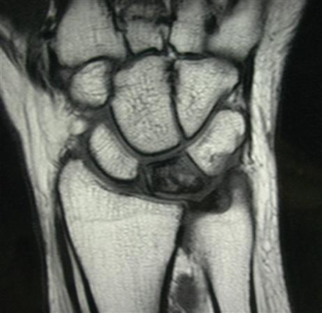
MRI scans are much more sensitive during the early stages because they can detect changes in the blood supply and bone before collapse occurs.
For this reason, MRI is often the most useful investigation when Kienböck’s disease is suspected.
Staging the Disease
Doctors often classify Kienböck’s disease into stages based on how much damage has occurred.
These stages describe whether:
- The blood supply has been affected
- The bone has started to collapse
- The surrounding wrist bones have become involved
- Arthritis has developed
The stage of the disease helps guide treatment decisions.
Treatment Options
Treatment depends on several factors, including:
- The stage of the disease
- Your symptoms
- Your age
- Your activity level
- Whether the bone has collapsed
- Whether arthritis is present
Non-Surgical Treatment
In the earlier stages, many people can initially be managed without surgery.
Activity Modification
Reducing activities that place high loads through the wrist can help reduce pain and slow further irritation.
This may include limiting:
- Heavy lifting
- Repetitive gripping
- High-impact activities
- Weight-bearing exercises through the hands
Wrist Splints
A wrist splint may be recommended to reduce stress on the lunate and allow symptoms to settle.
This is often used during painful flare-ups or in the early stages of treatment.
Pain Relief
Painkillers or anti-inflammatory medication may help improve comfort.
However, these medications do not restore blood supply to the bone or reverse the disease process.
Physiotherapy
Physiotherapy can help improve wrist movement, maintain strength, reduce stiffness, and guide a gradual return to activity.
It is important to understand, however, that physiotherapy cannot reverse bone collapse if significant structural damage has already occurred.
Surgical Treatment
If symptoms continue despite conservative treatment, or if imaging shows progression of the disease, surgery may be recommended.
The type of surgery depends on the stage of the disease and the condition of the wrist.
Joint Levelling Procedures
For people with negative ulnar variance, surgeons may alter the length of the forearm bones to reduce pressure on the lunate.
This may involve shortening the radius or, less commonly, lengthening the ulna.
The aim is to redistribute the forces passing through the wrist.
Vascularised Bone Grafts
Some procedures attempt to restore blood supply to the lunate using a bone graft that carries its own blood vessels.
These operations are known as vascularised bone grafts and are generally considered in selected patients with earlier-stage disease.
Salvage Procedures
If the lunate has collapsed and arthritis has developed, more extensive surgery may be necessary.
These procedures aim to relieve pain while preserving as much wrist movement as possible.
Examples include:
- Partial wrist fusion
- Proximal row carpectomy
- Other reconstructive procedures depending on the individual case
Although these operations cannot restore a completely normal wrist, they can often provide significant pain relief and improve function.
What Is the Outlook?
The outlook varies from person to person.
Some people remain relatively stable for many years with mild symptoms, while others experience gradual progression of the disease.
Early diagnosis generally provides more treatment options before significant collapse occurs, which is why persistent wrist pain should always be properly assessed.
With appropriate treatment, many people are able to continue working, remain active, and maintain good wrist function.
Key Points
- Kienböck’s disease affects the lunate, one of the small bones in the wrist.
- It occurs because the blood supply to the bone becomes reduced, leading to avascular necrosis.
- Symptoms commonly include wrist pain, stiffness, weakness and reduced grip strength.
- MRI scans are often needed to diagnose the condition in its early stages.
- Treatment ranges from activity modification and splinting to physiotherapy and surgery, depending on the severity of the disease.
- Early diagnosis and appropriate management offer the best chance of preserving wrist function.
This article is for general information only and does not replace individual assessment. If you’re concerned about your symptoms, please consult a qualified healthcare professional. See our full medical disclaimer.